Anatomy A215 Virtual
Microscopy
|
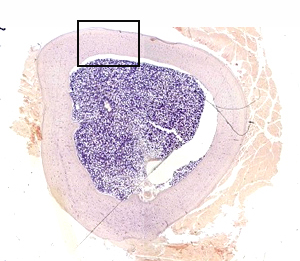 |
This is a cross section of bone with a central bone marrow cavity
containing red bone marrow. (Of course because of the staining for
microscopic analysis, the bone marrow is blue.) The section includes
a little muscle and soft tissue attached, but the elements of bone
should be quite apparent. The little box indicates where
the image below came from. |
|
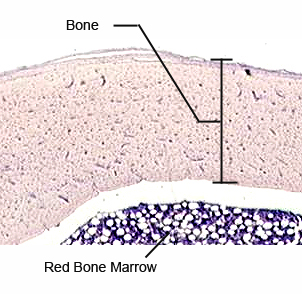 |
The features of bone you need to
identify on the DECALCIFIED BONE slide are:
- Central (or Haversian) canals
- Lacunae, containing osteocytes, between concentric layers of
bone
- Perforating (or Volkmann’s) canals, transversely connecting
central canals, and
- Lamellae (singular: lamella), the concentric layers of bone
between lacunae
Click here for an image
detailing these features.
|
A215 Home Page
| Virtual Microscopy
Table of Contents
|