Anatomy A215 Virtual Microscopy
Cochlea


The inner ear lies within the petrous part of the temporal bone and has two components: the outer bony (or osseouslabyrinth consisting of spaces in the bone; and the  membranous labyrinth suspended inside it.

Specifically, we will be examining the cochlea (part of the bony labyrinth) and the membranous structures within it. Identify the following:

  • The cochlear duct (or scala media), which splits the bony canal of the cochlea into the scala vestibuli and scala tympani.
  • The thicker basilar membrane, which separates the cochlear duct from the scala tympani.
  • The thinner vestibular membrane, which separates the cochlear duct from the scala vestibuli.
  • The spiral organ (or organ of Corti).

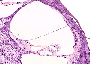
The details of the spiral organ may be seen under higher magnification. The spiral organ consists of hair cells that rest on the basilar membrane. The tectorial membrane is located in close proximity to the hair cells. Vibration of the hair cells (from sound waves) causes the stereocilia to bend against the tectorial membrane, producing a nerve impulse that is sent to the brain as sound.

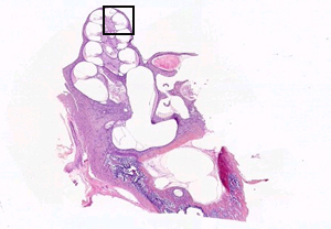
The image to the left is from the little black box above.

See this slide with the virtual microscope

I don't see the virtual slide, I must need the download.


   A215 Home Page | Virtual Microscopy Table of Contents