Anatomy A215 Virtual
Microscopy
|
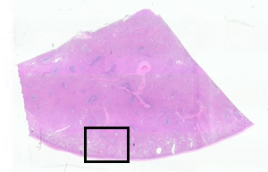 |
The spleen has a capsule and trabeculae, but lymphatic
vessels enter and leave the spleen only at its hilus, as do blood
vessels. There are many distinct regions classed as either red pulp
or white pulp according to their unstained appearance. The
difference is apparent at low magnification.The box in the image
to the left represents the area of magnification below.
|
|
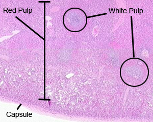 |
In this virtual microscopic slide of
SPLEEN, you should be able to identify:
- Red pulp, which is usually the more abundant. It consists of
numerous sinuses filled with red blood
cells.
- White pulp, which is found around the small blood vessels
which branch into the red pulp. White pulp typically consists of
a light staining central area near each vessel, surrounded by a
darker staining
dense aggregation of lymphocytes.
|
A215 Home Page
| Virtual Microscopy
Table of Contents
|