Microscopic features of the eye: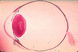
- Review the general organization of
the eye.
- Examine a section of the eye with low power and, from anterior to
posterior, identify
- The cornea,
- Iris,
- Lens,
- Ciliary body,
- Retina, and
- Optic nerve.
How does the cornea differ
histologically and functionally from the sclera?
Compare and contrast Bowman’s and Descemet’s membranes.
With
higher power, examine the retina carefully and note capillaries and
the ten layers.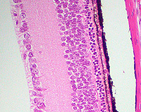
- Within these layers, identify the
pigmented epithelial cells, the rods and cones,
- The 3 layers of cell bodies, the
inner and outer plexiform layers, and the inner limiting membrane.
- Determine whether the fovea
or the optic disc (where the optic nerve meets incoming axons
from the retina is present on your slide and if not
share your neighbor's slide to observe these structures.
What must light pass through
before it hits rods and cones?
What exactly are the “photoreceptors” in the rods and cones?
What is the significance of the neuronal cell bodies in the retina?
Indicate three functions of the pigmented layer of the retina.
More about the
eye. |