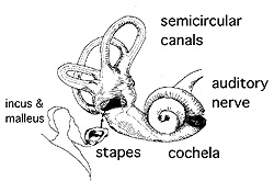
 The
Ear: A good understanding of the structures in the inner
ear requires that you be able to interpret the images you see under
the microscope in three dimensions. Since a single section will not
allow you to visualize these areas in different planes, it is
important for you to refer to the pictures in your texts to acquire
an appreciation for the three dimensional configuration of these
structures. The
Ear: A good understanding of the structures in the inner
ear requires that you be able to interpret the images you see under
the microscope in three dimensions. Since a single section will not
allow you to visualize these areas in different planes, it is
important for you to refer to the pictures in your texts to acquire
an appreciation for the three dimensional configuration of these
structures.
Clincial
note: Bacterial infections of the middle ear cavity (otitis
media) are common complications of colds and upper respiratory tract
infections in small children. If such an infection does not respond
to antibiotics, the resulting fluid and inflammatory material may be
drained through a perforation in the tympanic membrane. IN the image
to the right the ear drum on the left is healthy, whereas the one on
the right is markedly inflamed and must have hurt. may be
drained through a perforation in the tympanic membrane. IN the image
to the right the ear drum on the left is healthy, whereas the one on
the right is markedly inflamed and must have hurt.
Examine a section of the inner ear. This specimen was dissected from the skull and
sectioned to best demonstrate structures within the conical,
spiral-shaped cochlea. Identify
- Portions of the spiral ganglion and
spiral organ of Corti.
- Choose the best region showing the
organ of Corti and identify the associated ducts (the scala vestibuli, scala media, scala tympani),
- The vestibular membrane,
- Tectorial membrane, and
- The major sensory or hair cells.
About that ear
fluid. |