 Cells
in Lymph and the thymus Cells
in Lymph and the thymus
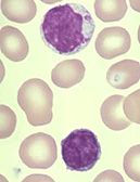
Lymphocytes in blood smear are shown in the image to the
right.
Thymus
First note the capsule and septa.
Next notice the organization of lobes into a basophilic cortex and
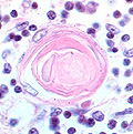
eosinophilic medulla. A key feature of the medulla is the presence
of Hassal’s corpuscles, which are masses of degenerated epithelial
cells. The epithelial reticular cells make up the framework of the
thymus.
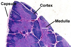
- The capsule,
- Septa, and
- The organization of lobes into a
basophilic cortex and eosinophilic medulla.
Identify the epithelial-reticular
cells or epitheliocytes which make up the framework of
the thymus.
- These are larger and paler than
the lymphocytes. In the
medulla,
identify the masses of epithelial cells, thymic or Hassal's
corpuscles.
Identify macrophages in
the thymic cortex (they are typically larger and more pale than
lymphocytes and less numerous than the epithelial-reticular cells) and look for mitotic figures among the lymphocytes of
this region.
What is the function of the
individual epithelial reticular cells?
How do the lymphocytes seen here differ
from those in the blood smears studied earlier?
Next let's look at a
lymph node. |