General
and Systemic Histopathology, C601&C602
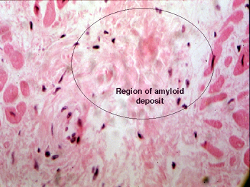
Slide 8: Heart with amyloid deposition
 |
It is difficult to recognize
the amyloid protein in this slide. It appears as an amorphous staining
pink or orange fibrillary material in between the myocytes that make up
the heart muscle. It may be easier to spot in the walls of the smaller
arteries that feed the myocardium. It shows up best with a "congo red"
stain. How do you think the addition of lots of non compliant protein in
between the muscle fibers would alter the action of the heart?
See this slide with the
virtual microscope. |
Back
to Home
|