
General
and Systemic Histopathology, C601&C602
Slide 76: Lung with tuberculosis
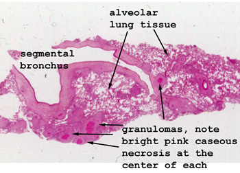 |
Look carefully at the
lung tissue for the little pink areas of caseous necrosis. These are the
areas of tubercular infection. They don't show a well developed granuloma
architecture, but you'll see the evolving features and should have no trouble
finding the giant cells.
See this slide with the
virtual microscope. |
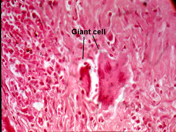 |
This picture details
two of the giant cells associated with one of the granulomas. You can see
the peripheral array pattern of the nuclei in the larger one. The age of
this slide has contributed to the "hot pink" staining quality. Other than
the accentuation of the eosinophilic property of the stain, this slide
is very good representation of the tubercular process. |
Back
to Home
|