General and Systemic Histopathology, C601&C602
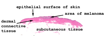
Slide 116: Skin with malignant melanoma

|
Although this is just
a little shave biopsy of skin, you can easily see the central area of thickening
where the melanoma is. See this slide with the virtual microscope. |

|
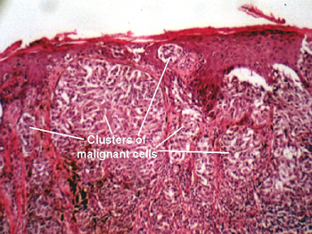
The skin in this slide shows clusters of malignant melanocytes in the dermis. Observe the lack of "cohesion" of the cells. Nuclear features of malignancy should be obvious, and many cells will show abundant pigment. There is no " maturation from surface to base " in this lesion, an important consideration in distinguishing this from its benign counter-part, a "nevus." Depth of penetration is a critical part of "staging" this lesion. |
Neoplasia Lab Page
| Previous Slide
| Next Slide
| Table of Contents
Mark
Braun, MD braunm@indiana.edu
Copyright 1998,
the Trustees of Indiana University