
General
and Systemic Histopathology, C601&C602
Slide 157: Skin with recurrent squamous
cell carcinoma
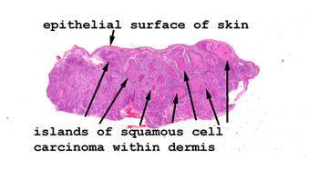 |
Here you see the groups
of malignant cells within the dermis but seemingly having no connection
to the epidermis. What's the explanation?
See this slide with the
virtual microscope. |
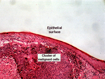 |
The picture of this slide
could be in focus a little better, but it's what we have. Note the epithelium
does not show changes of nuclear atypia nor cancer. The squamous cancer
is in the dermis, and represents a recurrence from a previously removed
malignancy. On your slide, you should be able to see the hallmark nuclear
features of cancer i.e. angulated nuclear margins, hyperchromasia and reduced
nuclear to cytoplasmic ratio. Look for "intracellular" bridges between
the malignant cells. |
Back
to Home
|