
General
and Systemic Histopathology, C601&C602
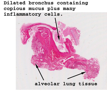
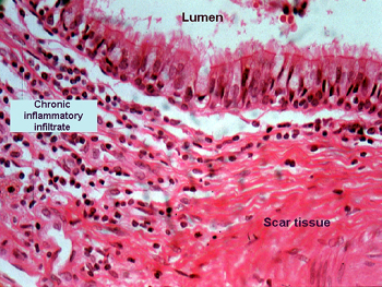
Slide 70: Lung with bronchiectasis
 |
Things to look for in
this slide include the amount of fibrosis and inflammation in the wall
of the bronchus as well as the mucus in the bronchial lumen.
What about the number of goblet cells in the mucosa.
See this slide with the
virtual microscope. |
 |
This condition is hard
to show on a microscopic slide. In bronchiectasis, the bronchi become dilated
and often fill with mucus, bugs and inflammatory debris. People with this
condition cough and bring up lots of sputum, and generally have a heck
of a time with infections. Sometimes the infections can spread via the
blood stream to other organs. Look in the wall of the bronchus in this
section and see the degree ofinflammation. It is hard to get the perspective
of a markedly dilated bronchus though. |
Back
to Home
|