| Your slide consists of
a very small strip taken from the wall of a very large cystic tumor.
Don't waste a lot of time trying to find normal ovary, I'm not sure there
is any.
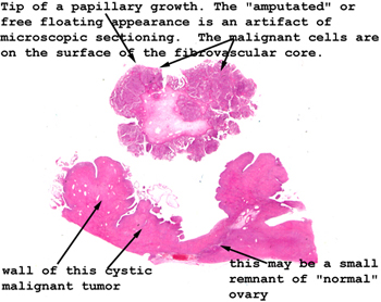
The malignant epithelium
is found lining the cyst and on the surface of the many papillary growths
and extensions. The papillary "head" seen in the picture to the left
appears to be "floating" because when the section was cut the
stalk connecting it to the wall of the cyst out of the plane of the section.
Trust me, it was connected to the wall.
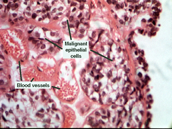
Look on the surface of
this little "head piece" for the best examples of the malignant epithelium.
See this slide with the
virtual microscope. |