
General
and Systemic Histopathology, C601&C602
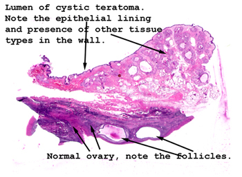
Slide 163: Ovarian teratoma
|
This picture nicely
shows a portion of the ovary compressed at the edge of the cyst. The
cyst lumen would be at the top of the picture. Note the many different
kinds tissue in the wall of the tumor. Remember, this a benign lesion.
See this slide with the
virtual microscope. |
|
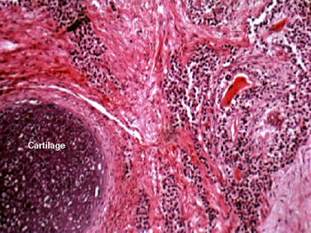
In this field you see
some mature cartilage intermixed with other elements. These lesions are typically
formed of benign tissue elements of all three embryonic germ layers. The
most common constituent is skin, including both epidermis and dermis, but
some of your slides include bowel and respiratory mucosa, and a few sections
even had cerebellum. This degree of tissue diversity is probably not surprising
given the origin of the tumor. Although these lesions are almost invariably
benign in females, the testicular counterpart in a man is generally malignant. |
Back to Home
|