
General
and Systemic Histopathology, C601&C602
Slide 7: Healing fracture of bone.
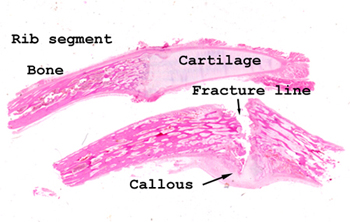 |
This slide obviously
has two sections of tissue. Both are of a rib and the lower portion shows
a partially healed fracture line. You can see the developing callous
on one side and there is abundant granulation tissue in the fracture line
itself. We split the fracture line open at the time the specimen
was embedded so as to highlight where to look for the healing process.
In life, the fracture was closed and the edges were knit together with
the newly formed granulation tissue.
See this slide with the
virtual microscope. |
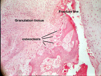 |
This healing "fracture"
is approximately two weeks old, and shows early changes of the healing
process. Note the remodeling taking place by the osteoclasts and the rather
marked degree of fibrosis (scarring) that is taking place as the new bone
is being formed. In your slide, you should be able to see many active fibroblasts
and angioblasts as part of the initial healing "team." See if you can find
the area just by looking at your slide. |
Back
to Home
|