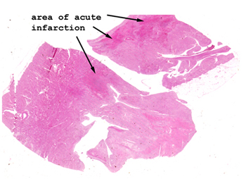
Here the infarct shows
areas of scar formation, but still you can see the typical coagulative necrosis
that typifies anoxic injury. The cells develop a glassy eosinophilic appearance,
eventually lose their nuclei, lyse and are removed by scavenger cells such
as monocytes. You should know the basic stages of the development of a myocardial
infarction, and what you would expect to see grossly and microscopically
at day 1, 3, 5, 7 and 10, assuming the patient lives that long. You must
also know what are the causes of "sudden" death with a myocardial infarction.
For example, between day five and seven, the infarct is the weakest and could
rupture. What to you suppose happens when the heart ruptures? No, surprisingly,
the patient does not bleed to death.