
General
and Systemic Histopathology, C601&C602
Slide 6: Myocardial infarction
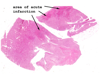 |
In this slide, look for
the areas of bright pink. They will represent the areas of acute
infarction. Can you place an age on this lesion?
See this slide with the
virtual microscope. |
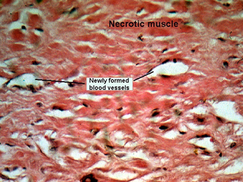 |
This is a high power
view of the edge of the infarct. Note the areas of in growth of new blood
vessels and the reactive nature of the fibroblasts. On low power, you will
be able to see the staining difference that indicates the regions of dead,
dying, dissolving and repairing myocardial tissue. This infarct is about
7 or 8 days old, but obviously the patient died. Had the person died instantly
at the time of the initial coronary occlusion, would
there be an area of infarction? |
Back
to Home
|