
General
and Systemic Histopathology, C601&C602
Slide 35: Retinal Melanoma
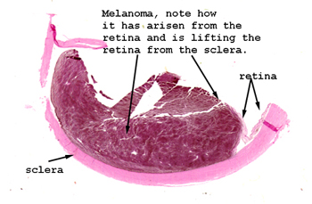 |
Again, there is probably
no trouble seeing the tumor in this slide. It is obviously black
because it is a melanoma. They can occur in various places in the
eye but this is probably most common.
See this slide with the
virtual microscope. |
 |
This slide shows a primary
malignant melanoma of the retina. Most of the histological features of
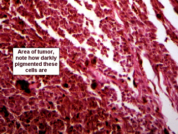
this tumor are just like those of cutaneous melanomas. You will note a
lot of pigment, so much so, in fact, that in many instances you won't be
able to see the nuclear morphology of the malignant cells. The one curious
feature of this tumor is its propensity to metastasize to the liver. I
have actually heard, jokingly so, the term "ocular-hepatic" shunt applied
to this property of retinal melanomas. For what it's worth, there are actually
three sites where primary ocular melanomas arise: the retina, the iris
and the conjunctiva. It is the retinal variety that frequently metastasizes
to the liver, and often very early in the course of the disease. |
Back
to Home
|