
General
and Systemic Histopathology, C601&C602
Slide 184: Glioblastoma Multiforme
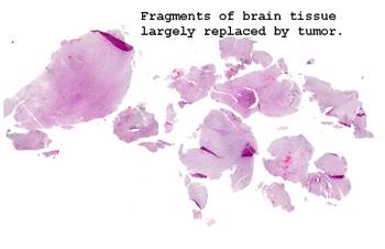 |
We see here just small
fragments of brain and tumor. This is how these specimens come to
us from surgery.
See this slide with the
virtual microscope. |
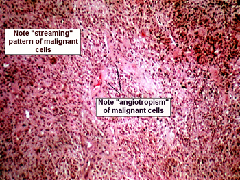 |
This is a high grade
malignant astrocytic glial tumor. You will see areas of necrosis and marked
nuclear atypia of the malignant cells. Pay special attention to the reactive,
yet highly atypical looking, vascular changes in the capillary sized vessels.
The endothelial cells become extremely agitated in the presence of this
tumor. You should see some large pink staining cells at the periphery of
this lesion; they are called gemistocytes, gem like cells. These are benign
reactive glial cells, and similar changes are seen in many types of central
nervous system injury. This tumor is highly aggressive and universally
fatal, at least at the present time. |
Back
to Home
|