
General
and Systemic Histopathology, C601&C602
Slide 24: Papillary Adenocarcinoma of the
Thyroid
|
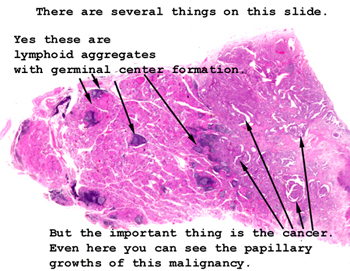
Note that you can actually
see the areas of papillary carcinoma just by looking at the tissue on the
slide. You will also see many lymphoid aggregates within the surrounding
thyroid tissue. This may reflect some overall excitement on the part
of the immune system or may indicate a coexisting thyroiditis.
See this slide with the
virtual microscope. |
|
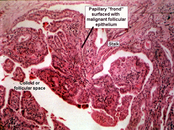
This picture pretty
well says it all. You will see papillary groups of fibrovascular tissue surfaced
with cuboidal or columnar epithelial cells. The epithelial cells covering
these papillary fronds are the malignant follicular cells. They perceive the
space between the papillary groups as the follicular lumen, although they
are not making much in the way of colloid. You will likely see some scarring
and a few chronic inflammatory cells in association with the tumor. |
Back to Home
|