
General
and Systemic Histopathology, C601&C602
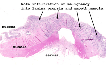
Slide 105: Adenocarcinoma of colon
|
Again, the picture
pretty well tells the story here. The malignancy has arisen from the colonic
mucosa and spread into the muscular wall. The large clear space in
the middle of this tissue is actually an artifact of the sectioning and is
not some change brought on by the tumor.
See this slide with the
virtual microscope.
|
|
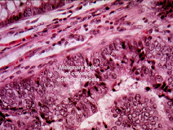
Again, looking at the
slide on a white background will help you see the area of malignancy. Microscopically,
look at a region where you can see both malignant and benign elements in the
same field. The difference in gland and cellular morphology will be much
more evident. The malignant glands show "gland within gland" pattern as well
as disorganized arborization and branching. The usual cellular features of
malignancy are here in abundance: nuclear-cytoplasmic ratio, hyperchromasia,
angulated nuclear margins etc. |
Back to Home
|