
General
and Systemic Histopathology, C601&C602
Slide 138: Stomach with signet ring
cell carcinoma
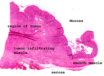 |
In this picture of the
tissue, you can see how greatly expanded the lamina propria and muscular
layers are. The type of cancer represented here is rather peculiar.
There may not be any evidence on the mucosal surface, yet there will be
extensive infiltration of the wall. The cells do not, as a rule,
organize themselves into gland-like structures, rather the diagnosis of
adenocarcinoma is made by seeing the large intracellular mucin vacuole
in each cell.
See this slide with the
virtual microscope.
|
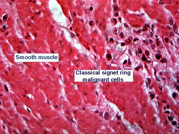 |
The changes here are
subtle. The tumor is not on the mucosal surface, but down in the muscle
and lamina propria. It may look like an inflammatory infiltrate spread
around between the bundles of muscle cells. Look for the hallmark features
of malignancy in the cytology of the cells. Only a few really diagnostic
signet ring cells will be present. These cells will have a large vacuole
off-setting the nucleus, thereby giving the "signet" ring appearance. |
Back
to Home
|