
General
and Systemic Histopathology, C601&C602
Slide 39: Esophagus with mucus cyst
|
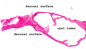
This slide shows a
benign mucinous cyst in the wall of the esophagus. First get yourself
oriented as to where the mucosal and the serosal surfaces are. Then
take note of the cyst lining.
See this slide with the
virtual microscope. |
|
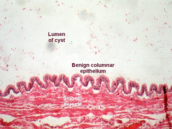
The picture in this
case shows only the cyst lining and a portion of the muscular wall of the
esophagus. Note that the lining of the cyst is columnar and mucous secreting
epithelium. That's about it for this slide. |
Back to Home
|