
General
and Systemic Histopathology, C601&C602
Slide 9: Stomach with gastric ulcer.

|
This picture actually
represents about half of the tissue on your slide. Just imagine it's
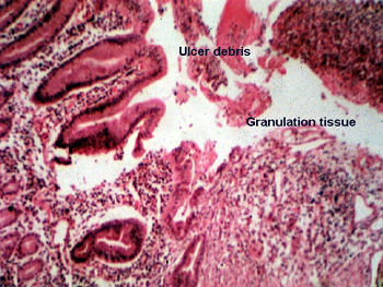
got a mirror image right half and you'll have it. The photomicrograph below
comes from the edge where the ulcer meets the mucosa. Note the bright
pink layer of fibrin and digested protein material that lines the ulcer base.
The granulation tissue begins just beneath this. See how the ulcer has eroded
completely through the muscle and is about to perforate through the serosal
fat. What do think is the significance of the what is seen just to
the left of the ulcer base?
See this slide with the
virtual microscope. |
|
Your slide includes
only about half the ulcer. Before going to the microscope, hold the slide
up to the light or look at it on a white background. You should be able to
easily spot the area of the ulcer. Try to get yourself oriented before diving
in with the scope. Observe the "granulation tissue" in the ulcer base and
be sure you can identify angioblasts and the numerous reactive fibroblasts.
There is considerable digested debris on the surface of the ulcer, don't confuse
this for the reparative elements of granulation tissue. The digested junk
contains epithelial cells, inflammatory elements, bacteria and who knows
what all. What's the bacterial agent
of such renown in this disease? |
Back to Home
|