
General
and Systemic Histopathology, C601&C602
Slide 110: Artery with atherosclerosis
and thrombosis
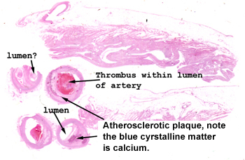 |
Spotting the vessels
with the atherosclerosis, narrowed lumens and thrombi should not be to
much of a challenge. Look at how compromised the lumen is even in the absence
of the thrombus. What you see here are multiple serial sections of
the same coronary artery.
See this slide with the
virtual microscope. |
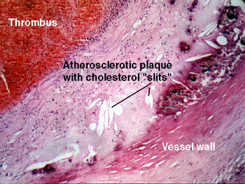 |
Although this condition
will be dealt with in detail in various sections of this course, here's
a chance for you to study the basic morphology of the plaque for its own
sake. This slide is of an elastic artery with a classical atherosclerotic
plaque with secondary thrombosis. The plaque is in the sub-intima and is
a fairly complex structure. Observe the cholesterol "slits." The cholesterol
was "washed out" during the processing of the tissue, leaving behind the
little spaces where the deposits had been. As far as problems associated
with this disease, a plaque can weaken the wall of an artery, potentially
causing a rupture of the vessel; it can cause thrombosis (as it did here)
and thereby complete occlusion of the lumen; and it can continue to "grow."
What do you think happened to the patient that gave us this slide? |
Back
to Home
|