
General
and Systemic Histopathology, C601&C602
Slide 114; Splenic Infarction
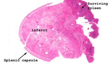 |
This picture shows
you the business end of this slide. The infarct represents most of
the tissue. I suggest you start at the edge of the normal tissue and
move into the area of the infarction.
See this slide with the
virtual microscope. |
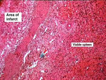 |
The picture pretty
well says it all here. You should be able to find the area of infarction
just by looking at the slide on a white background. Seeing this much hemorrhage
in a splenic infarct is a little unusual as the arterial supply is what we
refer to as an "end organ" type. One typically sees an anemic (white) infarct
in the spleen, but in this particular case there is some hemorrhage. Why do
you think this is so? |
Back
to Home
|