
General
and Systemic Histopathology, C601&C602
Slide 201: Lung with emphysema, bronchial
obstruction and pulmonary edema
 |
Even at low power, you’ll be able to see
the enlarged alveolar air sacs and mucus plugging of the airways.
See this slide with the
virtual microscope. |
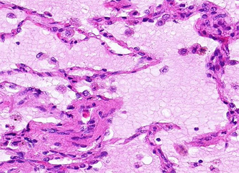 |
Here we see tremendous enlarged alveolar
spaces, filled with pink staining edema fluid. Many of the alveolar lining
cells have died and shed into the fluid filling these air spaces. The cells
peppered around in the edema are not inflammatory cells. You will, however,
see some chronic inflammation in the interstitial tissue and around the
vessels and airways. |
Back to Home
|