
General
and Systemic Histopathology, C601&C602
Slide 76: Lung with tuberculosis
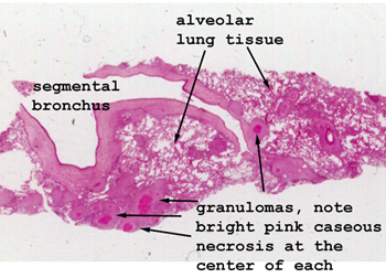 |
Look carefully at the
lung tissue for the little pink areas of caseous necrosis. These are the
areas of tubercular infection. They don't show a well developed granuloma
architecture, but you'll see the evolving features and should have no trouble
finding the giant cells.
See this slide with the
virtual microscope. |
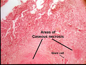 |
This section of lung
is largely replaced with a single granuloma. Note the structure of the
granuloma and the fact that it has a "caseous" center. There are numerous
giant cells and the fibrous margin of the granuloma is only partially formed
at this time. The caseous center of the granuloma is very characteristic
of TB. The granuloma itself, and the giant cells, represent a general reaction
to an agent the body cannot eliminate or destroy. |
Back
to Home
|