
General
and Systemic Histopathology, C601&C602
Slide 106: Syphilitic aortitis
|
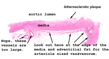
You have to use your
imagination a bit here. The tissue you have is just a little strip
out of a large aneurysm of the thoracic aorta. There is no way a complete
section would fit on your slide. So, the first thing to do is to get yourself
oriented as to where the lumen of the aorta is and where the adventitial
surface is. The atherosclerosis here is an incidental finding and not
necessarily part of the reaction to the treponemal bugs. After all,
this person is entitled to more than one disease.
See this slide with the
virtual microscope. |
|
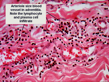
The inflammatory changes
in the wall of the aorta are subtle and observed only in the outer layers
of the muscular wall and the adventitia. The organism causes an obliterative
"end arteritis," and what you see is the chronic inflammatory infiltrate surrounding
the vasa vasorum. It is these smaller vessel that become the "target organ"
of the treponemal bug. The H&E stain does not stain the organisms. It
would take a special silver stain to highlight the typical corkscrew pattern
of this pathogen. Even with this stain, they would be hard to find, as so
few organisms are present. |
Back to Home
|