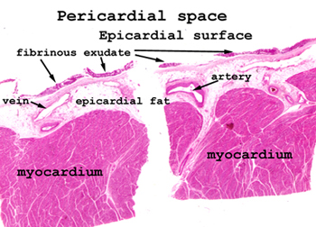
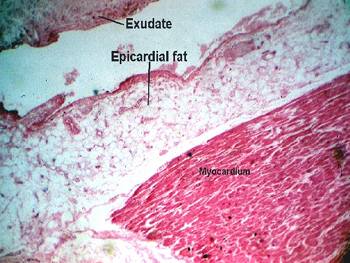
In this case, we're
looking for a thin band of homogeneous, pink staining, proteinaceous material
on the epicardial surface of the heart. This represents an exudate
composed largely of protein material. You will see very few inflammatory
cells.
Before putting slide
on the microscope stage, look at the tissue to find the epicardial surface.
This is where you will find the exudate. This exudate is almost totally devoid
of inflammatory cells, and consists almost totally of protein (fibrin plus
other trash). It looks the way I think "tofu" would look if sectioned and
stained. This exudate is a product of renal failure, is completely sterile.
It occurs secondary to the crystallization of nitrogenous wastes on the epicardial
and pericardial surfaces. Renal failure is consequence of many forms
of long term kidney disease.