
General
and Systemic Histopathology, C601&C602
Slide 126: Gallbladder with acute and
chronic inflammation
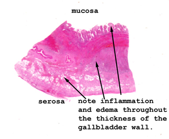 |
It's pretty obvious how
markedly thickened and edematous the wall of this gallbladder is.
Note the extensive inflammation throughout all layers of the wall.
See this slide with the
virtual microscope. |
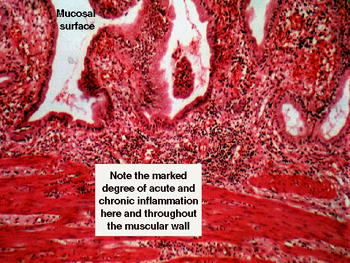 |
Are there ever numbers
of acute inflammatory cells in the wall of this gallbladder! You will see
a mixed infiltrate with both acute and chronic features. What defines the
two different patterns? Look at the mucosa and the full thickness of the
wall to get some idea of how inflamed this organ is. What are some of the
causes and consequences of this condition? |
Back
to Home
|