
General
and Systemic Histopathology, C601&C602
Slide 27: Gallbladder with acute and
chronic inflammation
 |
This section represents
only a small slice out of a dilated, inflamed (and no doubt painful) gallbladder.
Undoubtedly there were stones present as well, but we don't have any direct
microscopic evidence for them. Find the mucosa and then work your
way through the wall to the serosa. Pay attention to the inflammatory
cells and where you see them. What about the lamina propria?
See this slide with the
virtual microscope. |
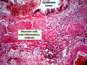 |
Find the lumen and try
to have yourself oriented before looking for the infiltrate. You will see
a mixed inflammatory infiltrate consisting of both "acute" and "chronic"
inflammatory cells, again lymphocytes are to be expected in the submucosa
of a structure associated with the gastrointestinal system. You will see
a large amount of granulation tissue on the serosal surface. |
Back
to Home
|