
General
and Systemic Histopathology, C601&C602
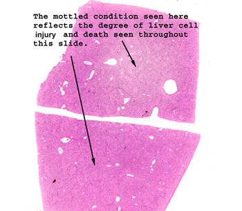
Slide 40: Liver with acute yellow atrophy
|
Again, the mottled
appearance of the tissue tells you there is some diffuse and generalized
process at work. I advise you to work your way down starting with the
lowest power of magnification and see if you can identify anything that looks
like a lobular pattern. Then go for high power and see if you can spot
triads and figure out what happened here.
See this slide with the
virtual microscope.
|
|
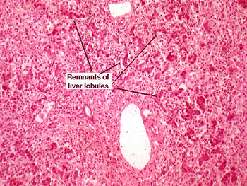
Acute Yellow Atrophy
is an old term, and not used much anymore. In this slide, so many hepatocytes
have died and been removed, that it is hard to tell this is even liver. Most
of what remains are bile ducts and triadal remnants. There is considerable
inflammation and absolute absence of the usual lobular arrangement. If you
are having trouble with this slide, you're in the majority; don't get too
worried. This resulted from a toxic exposure of chloroform, but many other
industrial volatiles can cause this same change. Clearly, this was an autopsy
specimen. |
Back to Home
|