
General
and Systemic Histopathology, C601&C602
Slide 47: Liver with metastatic cancer
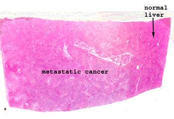 |
As with slides of this
sort, look at the uninvolved liver first and then move to the region of
pathology. The metastatic focus is pretty easy to recognize.
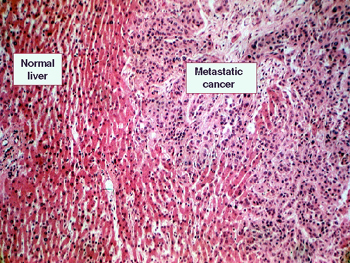
See if you can detect a glandular or "Indian file" pattern.
See this slide with the
virtual microscope. |
 |
Again, looking at this
slide on a white background will show the areas of cancer quite nicely.
I believe this is an example of metastatic breast cancer. You will see
rudimentary attempts to form glands by the malignant cells. Observe the
advancing margins of the tumors. Compare the cytology of the foreign malignant
cells to that of the surrounding healthy liver cells. Do you see vacuoles
in the metastatic malignant cells? |
Back
to Home
|