
General
and Systemic Histopathology, C601&C602
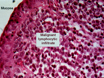
Slide 167: Intestinal Lymphoma
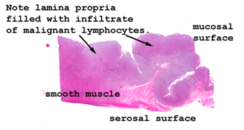 |
It's a lot easier to
see the changes in this slide than in #142. Here you can actually
see the infiltrative pattern of the tumor.
See this slide with the
virtual microscope. |
 |
This is the second example
of an intestinal lymphoma. The features are essentially the same as in
slide 142. See which is the better example in your set and spend time with
that one. You will see the malignant lymphoid infiltrate in the submucosa
and possibly extending through the muscular wall. Note the monomorphic
nature of the malignant lymphocytes. The distinction of nodular and diffuse
does not apply here; these terms only have reference in lymph nodes themselves. |
Back
to Home
|