
General
and Systemic Histopathology, C601&C602
Slide 32: Adenocarcinoma of rectum
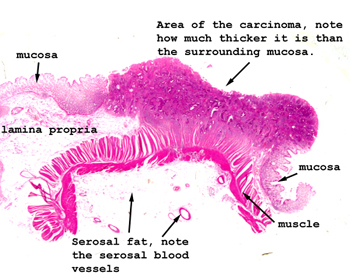 |
Here the region of the
tumor is pretty obvious. Look to see how it is spreading at the lateral
and deep margins. If we assume no node or distant metastasis what would
the Dukes classification of this lesion be? What of the TMN classification?
See this slide with the
virtual microscope.
|
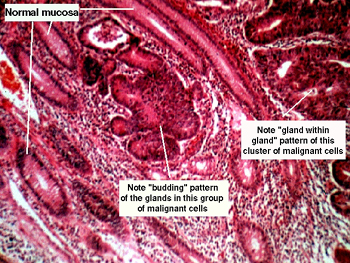 |
This is a fairly high
power view of the cancer with normal tissue at the edges. On low power,
you should be able to readily spot the different types of mucosa. In the
area of the cancer, observe the "branching and arborizing" gland margins
as well as the "gland within gland" pattern of the malignant cells. See
what we mean by "nuclear atypia" of the epithelial cells. They are hyperchromatic
with irregular nuclear staining and "angulated" nuclear margins. Mitoses
are every place. Note the spread into the lamina propria of the malignant
cells. Can you think of conditions that are associated with an increased
incidence of this condition? |
Back
to Home
|