
General
and Systemic Histopathology, C601&C602
Slide 79: Kidney with chronic pyelonephritis
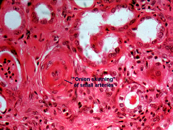 |
A higher power view showing
one of the more important vascular changes often associated with this condition.
These are small vessel changes of hypertension, which speak to the relative
degree of hypoxia of the renal tissue. As noted in the first picture of
this condition, you will see many chronic inflammatory cells in the interstitial
tissue and dilated tubules containing pink staining proteinaceous goo,
giving the appearance of thyroid colloid. You should note the scaring in
the interstitial tissue in general and to some degree around the around
the glomeruli. This is often associated with chronic ischemic injury of
the kidney, which worsens as the process proceeds. Think diabetes and hypertension.
See this slide with the
virtual microscope. |
Back
to Home
|