
General
and Systemic Histopathology, C601&C602
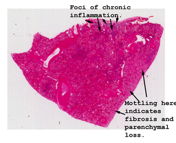
Slide 79: Kidney with chronic pyelonephritis
|
Even with no
magnification you can see the scattered blue staining that represents the
large number of lymphocytes in the interstitial tissue. It is possible
that you may be able to see little bright pink globs that represent the dilated,
protein containing tubules.
See this slide with the
virtual microscope. |
|
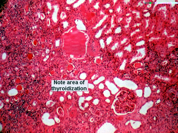
Note here the changes
we call "thyroidization." You will see many chronic inflammatory cells in
the interstitial tissue and dilated tubules containing pink staining proteinaceous
goo, giving the appearance of thyroid colloid. You should note the scarring
in the interstitial tissue in general and to some degree around the glomeruli.
This is often associated with chronic ischemic injury of the kidney, which
worsens as the process proceeds. Think diabetes and hypertension. |
Back to Home
|