
General
and Systemic Histopathology, C601&C602
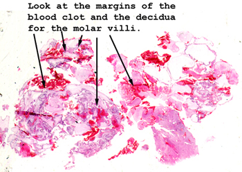
Slide 155: Hydatidiform mole
|
Look in the blood
clot or at the margin of the clot and decidualized endometrium for these bizarre
villi. You might want to compare these placental villi with those of
the first trimester miscarriage in slide 94. There is quite a difference.
Take note of the changes in the trophoblasts covering the villi.
See this slide with the
virtual microscope. |
|
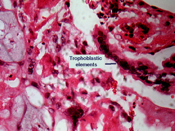
This is a higher power
view of the trophoblastic element of this "molar pregnancy." Note the large
and abnormally shaped villi with edematous cores. These villi are covered
with atypical trophoblastic cells growing as a syncytium. You may see a mitotic
figure or two, but on the whole, the degree of anaplasia is not nearly as
great as seen in a choriocarcinoma, the highly aggressive malignant counterpart
of this lesion. There will be some necrosis and inflammatory debris mixed
with the blood clot, but for the most part this is well preserved and very
representative. |
Back to Home
|