
General
and Systemic Histopathology, C601&C602
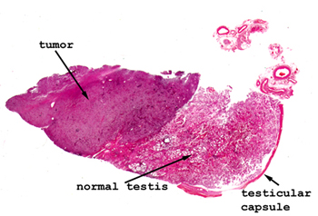
Slide 162: Testicular seminoma
|
This scan shows quite
nicely the area of tumor with the normal testis at the edge. See if
you can recognize a tubular arrangement of the malignant cells. There
will be a rather marked lymphocytic infiltrate as well. Sometimes this
lymphocytic element is so pronounced that these tumor are occasionally mistaken
for lymphomas.
See this slide with the
virtual microscope. |
|
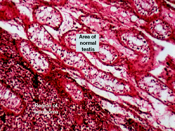
This picture shows
mostly normal testicular parenchyma with only a little tumor at the edge.
Note the "watery" appearance of the cytoplasm of the malignant cells and
their slight off center vesicular, "fried egg" looking nucleus. You may see
the cells in little clusters something like seminiferous tubules. You will
see the cells are very monotonous and bear some resemblance to lymphocytes.
Because there is often a significant lymphocytic infiltrate along with this
lesion, it is sometimes hard to distinguish from a testicular lymphoma. You
will see some mitoses, and possibly some embryonic tissues mixed with the
seminomatous elements. |
Back to Home
|