
General
and Systemic Histopathology, C601&C602
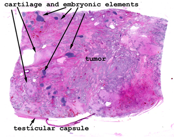
Slide 178: Mixed lineage carcinoma of testis
with elements of teratoma, seminoma and embyronal carcinoma
|
Even without any magnification
you can see the tremendous variation in tissue types in this slide.
I am not too sure you'll find much in the way of normal testis.
See this slide with the
virtual microscope. |
|
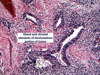
Note the multiple tissue
types in this tumor. The picture reveals several gland-like structures lined
by a columnar epithelium. There is an intervening stromal material composed
of mesenchymal elements that look very embryonic and undifferentiated. This
"embryonic" appearance gives rise to this lesion's name. You will see some
mitoses and areas of necrosis with accompanying inflammation. In some of
the slides, elements of a seminoma are also present. |
Back to Home
|