
General
and Systemic Histopathology, C601&C602
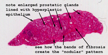
Slide 85: Prostatic hyperplasia
|
Here you can easily
see how the prostatic tissue is expanded with the mucin secreting glands.
Note the fibrous tissue septal divisions. You are likely to see a number
of lymphocytes within the stroma.
See this slide with the
virtual microscope. |
|
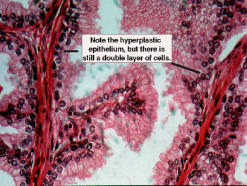
Hyperplasia of prostate,
high power view. Note the serrated borders of the prostatic glands with the
"piled up" and hyperplastic epithelial covering. You should be able to see
a DOUBLE layer of epithelial cells in all the glands and there should be
no mitoses. You will likely see some chronic inflammation in the interstitial
tissue. Remember, this slide is almost certainly from an old man, and more
than one condition may be present. |
Back to Home
|