
General
and Systemic Histopathology, C601&C602
Slide 146: Testicular lymphoma
|
This slide just all
malignant lymphocytes and I don't think there is anything even resembling
testis on this slide. No, I'd never give you something like this as
a quiz slide. Do the best you can with it.
See this slide with the
virtual microscope. |
|
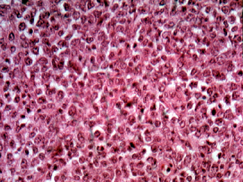
It would be impossible
to tell the tissue source from the picture I have taken. Note the uniform
infiltrate of monotonous lymphocytes throughout the entire specimen. Again,
the object here is to be sure this tumor is not an embryonal carcinoma or
version of seminoma, two common primary lesions of the testis. The treatments
are completely different. Use this slide for comparison when studying the
other testicular tumors we are about to see. |
Back to Home
|