
General
and Systemic Histopathology, C601&C602
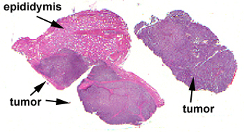
Slide 177: Seminoma of testis
|
In this slide there
isn’t a lot to tell you what the organ is. The epididymis should be recognizable,
but I doubt there is any identifiable testicular tissue. Most of the section
consists of the seminoma.
See this slide with the
virtual microscope.
|
|
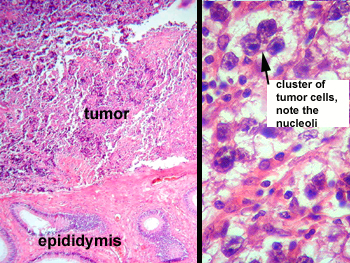
This split frame image
pretty well shows it all. The tumor cells appear as very immature cells that
one might see in the basal most layer of the seminiferous tubules. The nuclei
are extremely large and many have gigantic nucleoli. The tumor cells are
seen in clusters and you should see many lymphocytes within the tumor and
the bands of fibrous tissue that divide it.
Where else can
seminomas develop?
|
Back to Home
|