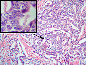
| This section nicely
shows the famous “gland-within-gland” pattern of the adenocarcinoma. The
insert shows the cellular detail from the edge of one of these malignant
gland forms. Note the angulation of the nuclear membranes and prominent nucleoli.
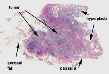
Be sure to check
the capsule of the gland for invasion and pay attention to the little nerves
in the surrounding fat. I don’t recall seeing perineural invasion, but it
might be present on your slide.
What about PSA?
Would you expect it elevated in this case?
|