
General
and Systemic Histopathology, C601&C602
Slide 150: Intradermal Nevus of Skin
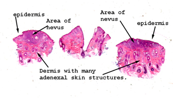 |
Again, it's fairly easy
to spot the lesion just by looking at the slide before going to your microscope.
You will see a number of hair shafts and sebaceous glands, some of which
are bit deformed due to the presence of the nevus. Even so, this
lesion is still benign.
See this slide with the
virtual microscope. |
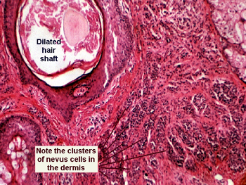 |
In this case you will
see clusters and theques of benign nevus cells in the dermis. You should
be able to see there is "maturation" of the cells as one goes from the
surface of the lesion to the base. You will find no mitosis and there is
no cytoatypia of the nevus cells. How would you distinguish this lesion
from a malignant melanoma? |
Back
to Home
|