
General
and Systemic Histopathology, C601&C602
Slide 151: Seborrheic Keratosis of
Skin
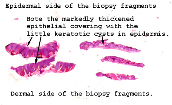 |
It's not real easy to
see what we're looking for in this biopsy, although once you recognize
it, you'll be surprised how abnormal the epidermis can look. The
thickening of the epidermis involves pretty much the whole surface.
This is a very common skin abnormality.
See this slide with the
virtual microscope. |
 |
This may fool you by
looking a bit like cancer, but it's not. This is a very common lesion.
You have seen them very likely as dark brown greasy looking spots on the
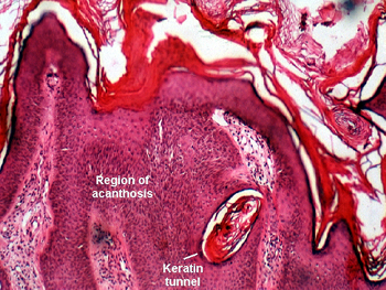
face, and especially around the temple, of older people. Note the hyperkeratosis
and acanthosis, that is to say the thickening and hyperplasia of the acanthotic
region and keratotic layers of the epidermis. You will also see what appears
to be little keratin inclusions cysts in the thickened epidermis. These
are often referred to as "pseudohorn" cysts and keratin tunnels. Even though
this lesion may look a little spooky, it is really not. Why do you suppose
this is not considered malignant? |
Back
to Home
|