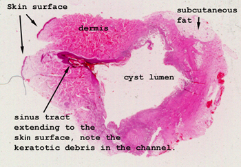
Here it's easy to understand
the nature of this lesion. We have a benign cyst, lined by stratified
squamous epithelium, present in the dermis. This probably came into
being as a puncture wound in the skin which drove a small piece of epidermis
down into the dermis. Rather than dying and being removed, it survived
and formed the cyst. In this case we even see a little sinus tract
communicating to the skin surface.
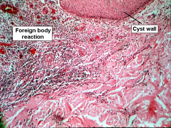
Note the cyst lining
composed of keratinized squamous epithelium. You should be able to find
the foreign body reaction at the edge of the cyst, and may even be able
to discern the site of rupture of the cyst wall. What are the giant cells
reacting to? How would a cyst of this type form? Try
slide 152 for another example, and answers to these questions.