Simple columnar epithelial cells
with cilia are numerous in the lining of the oviduct (slide 19),
where they are interspersed with secretory cells.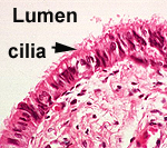
- Compare the appearance of the
cilia in this epithelium with the striated border (microvilli)
of absorptive epithelium on the last slide (slide 4).
- Examine the ultrastructural
views of the cilia and
microvilli shown in cross
section and in longitudinal section in Fig. 4-10. Compare cilia
to the absorptive stereocilia at the apical ends of the columnar
cells lining the epididymis (slides 39 and 40).
- The image to the right is of oviduct mucosa. Note the numerous cilia.
Answer this: How do cilia,
sterocilia and microvilli differ structurally and functionally?
Learning about this is not just an
academic exercise, rather it has real clinical significance.
-
Kartagener’s disease is an
inherited disorder involving mutations in the gene for dynein or
one of the many other proteins in cilia. All the cilia in such
individuals are immotile, leading to infertility and chronic
respiratory disorders.
How about
pseudostratified and stratified epithelium? |