 Larynx
begins the lower respiratory tract
(Fig. 17-1) and is closed by the epiglottis which prevents inspired air from
entering the esophagus and food/fluid from entering the trachea;
permits production of sounds. Larynx
begins the lower respiratory tract
(Fig. 17-1) and is closed by the epiglottis which prevents inspired air from
entering the esophagus and food/fluid from entering the trachea;
permits production of sounds.
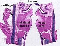
Slide 7 has a
longitudinal section extending through the larynx into trachea. The
section is similar to the right side of the example
shown here and in Fig. 17-4.
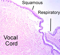
Examine the epithelium along this
tissue
- Note the change from stratified
squamous to pseudostratified, ciliated (or respiratory
epithelium).

- The protrusion with the large
mass of muscle represents one of the true vocal cords and the
protrusion that consists mostly of areolar support tissue is an
edge of a false vocal cord.
- Note the cartilage plates and
skeletal muscles of the larynx and the thyroid gland tissue
where the trachea begins.
What type of cartilage is found
in the larynx?
What histological differences
occur between the true and the false vocal cords?
Now for the
trachea. |