|
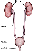 Ureter
-- tube continuous with the renal pelvis which transports urine
from kidney by peristalsis (Fig. 19-15). Ureter
-- tube continuous with the renal pelvis which transports urine
from kidney by peristalsis (Fig. 19-15).
Examine a section of ureter (slides
16 and
48), noting the highly
folded mucosa, surrounded by two layers of smooth muscle and
adventitia (Fig. 19-15b). In the folds of the urinary epithelium (or urothelium) note that the basement membrane is extremely thin and is
closely associated with capillaries.
How does urinary epithelium protect
underlying tissue from the toxic properties of urine?
Why is urinary epithelium not
required along the urethra lining?
Bladder -- collects urine from
ureters for drainage via urethra
Examine a Masson trichrome-stained
section of bladder (slide 12) and examine the folded urinary or
transitional epithelium and the collagen in the underlying lamina
propria (Fig. 19-16).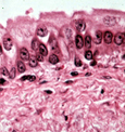
- Identify the three layers of
smooth muscle and the outer serosa/adventitia.
- The outer layer may contain
Pacinian corpuscles.
Why would Pacinian
corpuscles be located in the wall of the bladder?
What is the function of the
smooth muscle here?
Clinical note: Transitional cell carcinoma, the
most common type of bladder cancer, is associated with
occupational exposure to certain organic chemicals among workers
in the dye, rubber, paint, and some other industries. Smokers
also have a fourfold increased risk for bladder cancer compared
to nonsmokers. What
might explain the finding that cancer is more common in the
bladder than in the kidney or ureter?
Almost there, last comes
the urethra. |